
100%
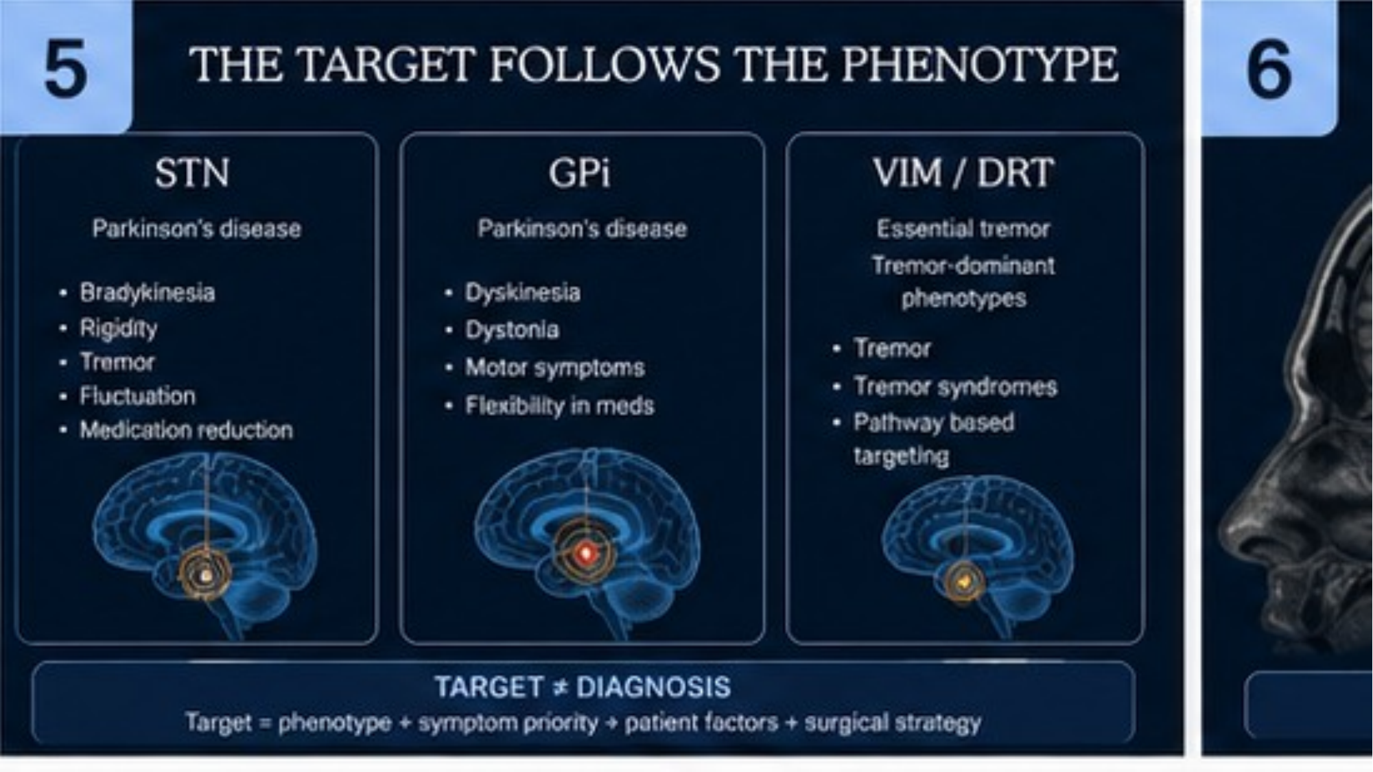
THE TARGET FOLLOWS THE PHENOTYPE
- STN
- Parkinson's disease
- Bradykinesia
- Rigidity
- Tremor
- Fluctuation
- Medication reduction
- GPi
- Parkinson's disease
- Dyskinesia
- Dystonia
- Motor symptoms
- Flexibility in meds
- VIM / DRT
- Essential tremor
- Tremor-dominant phenotypes
- Tremor
- Tremor syndromes
- Pathway based targeting
- TARGET ≠ DIAGNOSIS
- Target = phenotype + symptom priority + patient factors + surgical strategy
Image placeholder
THREE-TARGET ANATOMICAL INFOGRAPHIC
Real clinical image required
Image typeSchematic / diagram
DescriptionSimplified brain cross-section showing STN, GPi and VIM with subtle pale-blue target highlights; neighbouring structures labelled only where anatomically accurate.
Clinical purposeGive the three targets an anatomical location before stereotactic planning is introduced.
Suggested sourceConceptual anatomical diagram in the NEUROFLOW palette — clearly conceptual.
Copyright statusNot yet cleared
CaptionTo be written with the final image
AssociationModule 01 · Slide 05
Image placeholder
CORONAL BASAL GANGLIA ANATOMY
Real clinical image required
Image typeClinical MRI
DescriptionHigh-resolution anatomical MRI or atlas section showing STN, GPi, thalamus, internal capsule and substantia nigra.
Clinical purposeAnchor target discussion in real sectional anatomy rather than schematic approximation.
Suggested sourceProperly licensed stereotactic atlas or peer-reviewed publication.
Copyright statusTo be confirmed before publication.
CaptionTo be written with the final image
AssociationModule 01 · Slide 05
Image placeholder
VIM / THALAMIC ANATOMY
Real clinical image required
Image typeVIM anatomy
DescriptionAtlas section showing VIM relative to VPL, VPM, internal capsule and the subthalamic region.
Clinical purposeClarify why VIM stimulation side effects follow neighbouring thalamic and capsular anatomy.
Suggested sourceAuthoritative stereotactic atlas or appropriately sourced anatomical image.
Copyright statusTo be confirmed before publication.
CaptionTo be written with the final image
AssociationModule 01 · Slide 05