
100%
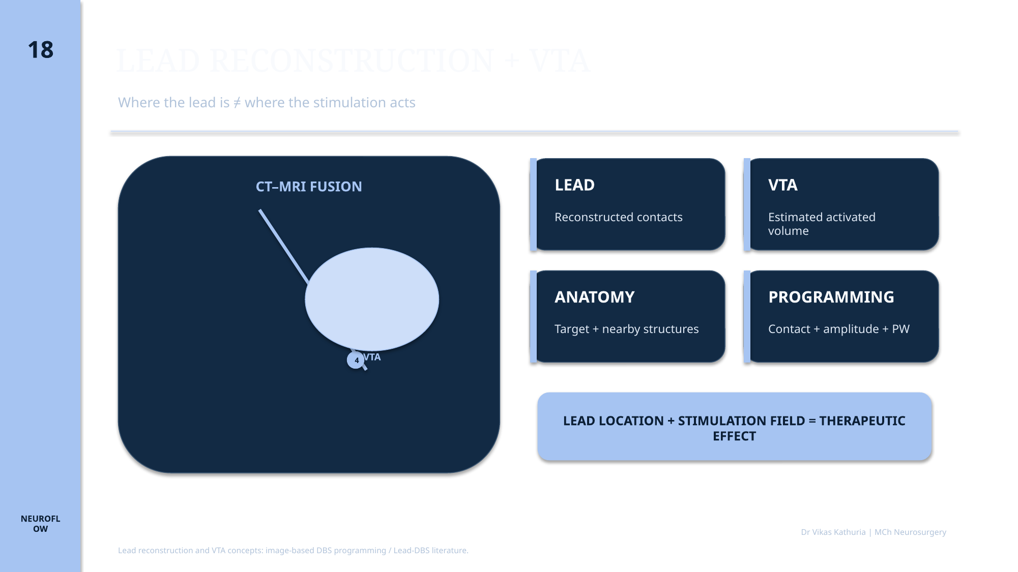
LEAD RECONSTRUCTION + VTA
- Where the lead is ≠ where the stimulation acts
- CT–MRI FUSION
- STN
- 1
- 2
- 3
- 4
- VTA
- LEAD — Reconstructed contacts
- VTA — Estimated activated volume
- ANATOMY — Target + nearby structures
- PROGRAMMING — Contact + amplitude + PW
- LEAD LOCATION + STIMULATION FIELD = THERAPEUTIC EFFECT
- Lead reconstruction and VTA concepts: image-based DBS programming / Lead-DBS literature.
- Dr Vikas Kathuria | MCh Neurosurgery
Image placeholder
18A POSTOPERATIVE CT WITH LEAD — INSERT AUTHENTIC IMAGE
Real clinical image required
Image typeCT
DescriptionPostoperative CT with lead
Clinical purposeSupport fellowship-level interpretation without fabricated clinical material.
Suggested sourceINSERT AUTHENTIC CLINICAL / PUBLISHED IMAGE HERE
Copyright statusSource and publication rights must be confirmed before use.
CaptionTo be written with the final image
AssociationModule 04 · Slide 18
Image placeholder
18B MRI–CT FUSION — INSERT AUTHENTIC IMAGE
Real clinical image required
Image typeMRI-CT fusion
DescriptionMRI–CT fusion
Clinical purposeSupport fellowship-level interpretation without fabricated clinical material.
Suggested sourceINSERT AUTHENTIC CLINICAL / PUBLISHED IMAGE HERE
Copyright statusSource and publication rights must be confirmed before use.
CaptionTo be written with the final image
AssociationModule 04 · Slide 18
Image placeholder
18C RECONSTRUCTED DBS CONTACTS — INSERT AUTHENTIC IMAGE
Real clinical image required
Image typePostoperative lead reconstruction
DescriptionReconstructed DBS contacts
Clinical purposeSupport fellowship-level interpretation without fabricated clinical material.
Suggested sourceINSERT AUTHENTIC CLINICAL / PUBLISHED IMAGE HERE
Copyright statusSource and publication rights must be confirmed before use.
CaptionTo be written with the final image
AssociationModule 04 · Slide 18
Image placeholder
18D VTA OVER STN — INSERT AUTHENTIC IMAGE
Real clinical image required
Image typeVTA visualization
DescriptionVTA over STN
Clinical purposeSupport fellowship-level interpretation without fabricated clinical material.
Suggested sourceINSERT AUTHENTIC CLINICAL / PUBLISHED IMAGE HERE
Copyright statusSource and publication rights must be confirmed before use.
CaptionTo be written with the final image
AssociationModule 04 · Slide 18
Image placeholder
18E VTA NEAR INTERNAL CAPSULE — INSERT AUTHENTIC IMAGE
Real clinical image required
Image typeVTA visualization
DescriptionVTA near internal capsule
Clinical purposeSupport fellowship-level interpretation without fabricated clinical material.
Suggested sourceINSERT AUTHENTIC CLINICAL / PUBLISHED IMAGE HERE
Copyright statusSource and publication rights must be confirmed before use.
CaptionTo be written with the final image
AssociationModule 04 · Slide 18
Image placeholder
18F VTA WITH TRACTOGRAPHY — INSERT AUTHENTIC IMAGE
Real clinical image required
Image typeTractography
DescriptionVTA with tractography
Clinical purposeSupport fellowship-level interpretation without fabricated clinical material.
Suggested sourceINSERT AUTHENTIC CLINICAL / PUBLISHED IMAGE HERE
Copyright statusSource and publication rights must be confirmed before use.
CaptionTo be written with the final image
AssociationModule 04 · Slide 18
Image placeholder
18G LEAD-DBS STYLE VISUALIZATION — INSERT AUTHENTIC IMAGE
Real clinical image required
Image typeVTA visualization
DescriptionLead-DBS style visualization
Clinical purposeSupport fellowship-level interpretation without fabricated clinical material.
Suggested sourceINSERT AUTHENTIC CLINICAL / PUBLISHED IMAGE HERE
Copyright statusSource and publication rights must be confirmed before use.
CaptionTo be written with the final image
AssociationModule 04 · Slide 18